0去购物车结算
购物车中还没有商品,赶紧选购吧!
当前位置: 图书分类 > 医药卫生 > 临床医学 > 肿瘤学 > Mammographic Image Analysis: Breast Cancer Risk Assessment and Computer-Aided Diagnosis

相同语种的商品

浏览历史

Mammographic Image Analysis: Breast Cancer Risk Assessment and Computer-Aided Diagnosis


联系编辑
 
标题:
 
内容:
 
联系方式:
 
  
Mammographic Image Analysis: Breast Cancer Risk Assessment and Computer-Aided Diagnosis
  • 书号:9787030665096
    作者:陈智丽等
  • 外文书名:
  • 装帧:平装
    开本:16
  • 页数:168
    字数:312000
    语种:zh-Hans
  • 出版社:科学出版社
    出版时间:2020-11-01
  • 所属分类:
  • 定价: ¥98.00元
    售价: ¥77.42元
  • 图书介质:
    纸质书

  • 购买数量: 件  商品库存: 6
  • 商品总价:

相同系列
全选

内容介绍

样章试读

用户评论

全部咨询

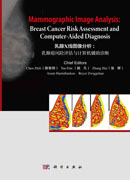
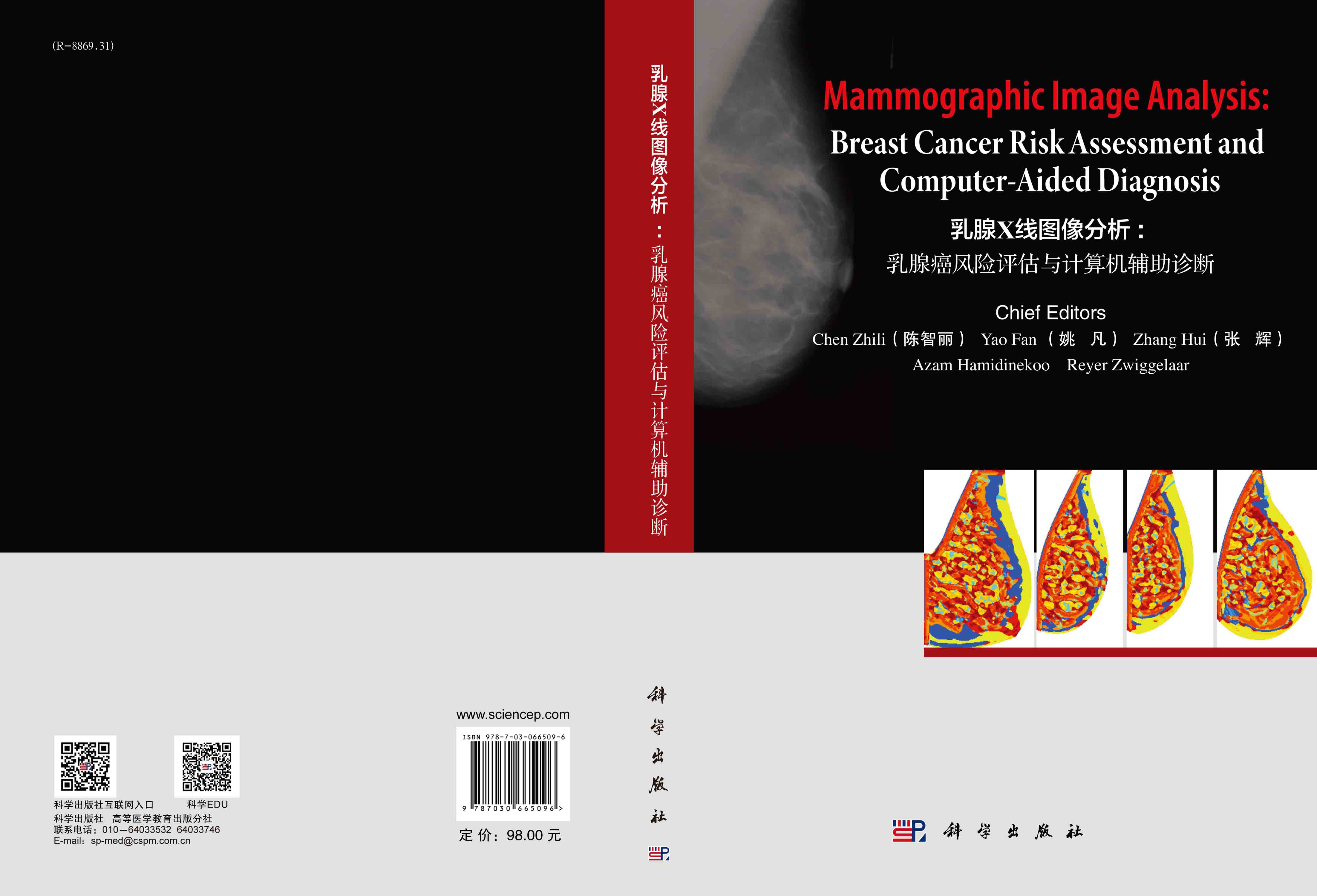
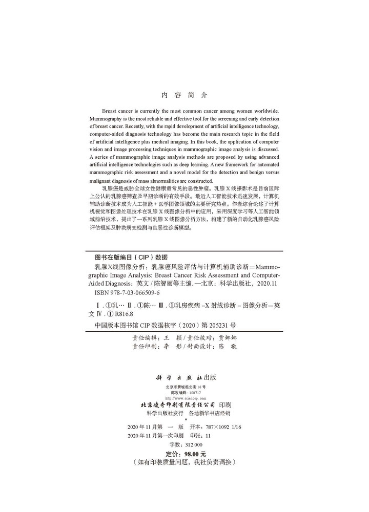
Breast cancer is currently the most common cancer among women worldwide. Mammography is the most reliable and effective tool for the screening and early detection of breast cancer. Recently, with the rapid development of artificial intelligence technology, computer-aided diagnosis technology has become the main research topic in the field of artificial intelligence plus medical imaging. In this book, the application of computer vision and image processing techniques in mammographic image analysis is discussed. A series of mammographic image analysis methods are proposed by using advanced artificial intelligence technologies such as deep learning. A new framework for automated mammographic risk assessment and a novel model for the detection and benign versus malignant diagnosis of mass abnormalities are constructed.
  乳腺癌是威胁全球女性健康最常见的恶性肿瘤。乳腺X线摄影术是目前国际上公认的乳腺癌筛查及早期诊断的有效手段。最近人工智能技术迅速发展,计算机辅助诊断技术成为人工智能+医学图像领域的主要研究热点。作者综合论述了计算机视觉和图像处理技术在乳腺X线图像分析中的应用,采用深度学习等人工智能领域前沿技术,提出了一系列乳腺X线图像分析方法,构建了新的自动化乳腺癌风险评估框架及肿块病变检测与良恶性诊断模型。
样章试读
  • 暂时还没有任何用户评论
总计 0 个记录,共 1 页。 第一页 上一页 下一页 最末页

全部咨询(共0条问答)

  • 暂时还没有任何用户咨询内容
总计 0 个记录,共 1 页。 第一页 上一页 下一页 最末页
用户名: 匿名用户
E-mail:
咨询内容:

目录

  • Contents
    Chapter 1 Introduction 1
    1.1 Breast Cancer Status 1
    1.2 Mammography 2
    1.3 Mammographic Risk Assessment 4
    1.3.1 Wolfe’s Four Risk Categories 4
    1.3.2 Boyd’s Six Class Categories 5
    1.3.3 Four BIRADS Density Categories 5
    1.3.4 Tabár’s Five Patterns 5
    1.4 CAD in Mammography 7
    1.5 Clinical Utility of the Present Research 8
    1.6 Focus and Contributions of the Book 8
    1.7 Book Outline 10
    Chapter 2 A Literature Review of Mammographic Image Analysis 12
    2.1 Mammographic Image Segmentation 12
    2.1.1 Breast Region Segmentation 12
    2.1.2 Breast Density Segmentation 19
    2.2 Estimation of Mammographic Density 23
    2.3 Characterisation of Mammographic Parenchymal Patterns 28
    2.4 Breast Density Classification 33
    2.5 Summary 37
    Chapter 3 Image Segmentation in Mammography 38
    3.1 Breast Region Segmentation in Mammograms 38
    3.1.1 Methodology 38
    3.1.2 Results and Discussion 42
    3.2 A Modified FCM Algorithm for Breast Density Segmentation 49
    3.2.1 FCM Algorithms 49
    3.2.2 A Modified FCM Algorithm 51
    3.2.3 Experimental Results 53
    3.3 Topographic Representation Based Breast Density Segmentation 57
    3.3.1 Topographic Representation 57
    3.3.2 Segmentation of Dense Tissue Regions 59
    3.3.3 Breast Density Quantification 61
    3.3.4 Results 62
    3.4 Summary 64
    Chapter 4 Texture Analysis in Mammography 66
    4.1 Local Feature Based Texture Representations 66
    4.1.1 Local Binary Patterns 67
    4.1.2 Local Grey-Level Appearances 67
    4.1.3 Basic Image Features 68
    4.1.4 Textons 69
    4.2 Mammographic Tissue Appearance Modelling 70
    4.3 Summary 74
    Chapter 5 Multiscale Blob Detection in Mammography 75
    5.1 Blob Detection 75
    5.1.1 Laplacian of Gaussian 75
    5.1.2 Difference of Gaussian 76
    5.1.3 Determinant of the Hessian Matrix 76
    5.1.4 Hessian-Laplacian 77
    5.1.5 Fast-Hessian 77
    5.1.6 Salient Region 77
    5.2 A Blob Based Representation of Mammographic Parenchymal Patterns 78
    5.2.1 Detection of Multiscale Blobs 79
    5.2.2 Blob Merging 85
    5.2.3 Blob Encoding 88
    5.3 Results and Discussion 88
    5.4 Summary 93
    Chapter 6 Breast Cancer Risk Assessment 95
    6.1 Experimental Data 95
    6.1.1 MIAS Database 95
    6.1.2 DDSM Database 96
    6.2 Evaluation Methodology 97
    6.2.1 Classification Algorithm 97
    6.2.2 Cross-Validation Scheme 98
    6.2.3 Result Representation 100
    6.3 Evaluating the Proposed Methods 100
    6.3.1 Evaluation of Breast Density Segmentation 100
    6.3.2 Evaluation of Breast Tissue Appearance Modelling 108
    6.3.3 A Combined Modelling of Breast Tissue 112
    6.3.4 Evaluation of Blob-Based Representation 115
    6.4 Summary 118
    Chapter 7 Discussions on Breast Cancer Risk Assessment in Mammography 120
    7.1 Comparison of the Proposed Methods 120
    7.2 Comparing with Related Publications 126
    7.3 Summary 130
    Chapter 8 Computer-Aided Diagnosis of Breast Cancer Based on Deep Learning 131
    8.1 Literature Review on Deep Learning Based Mammographic Image Analysis 131
    8.2 Mass Detection and Classification in Mammograms withaDeepPipeline 135
    8.2.1 Dataset Information 136
    8.2.2 Model Architecture 139
    8.2.3 Training 140
    8.2.4 Results & Discussion 140
    8.3 Summary 149
    Chapter 9 Conclusions 150
    9.1 Summary of the Book 150
    9.2 Contributions and Novel Aspects 152
    9.3 Future Work 154
    Bibliography 156
    Biography 167
帮助中心
公司简介
联系我们
常见问题
新手上路
发票制度
积分说明
购物指南
配送方式
配送时间及费用
配送查询说明
配送范围
快递查询
售后服务
退换货说明
退换货流程
投诉或建议
版权声明
经营资质
营业执照
出版社经营许可证